Anatomy Of Chest : Normal Anatomy of the Chest | Radiology Key : The chest can be split into two parts;. Use the mouse scroll wheel to move the images up and down alternatively use the tiny arrows (>>) on both side of the. Prep for a quiz or learn for fun! Fill out your shirt with a bigger, stronger, more powerful chest. It describes the theatre of events. There are two camps when it comes to chest training.
The thorax or chest is a part of the anatomy of humans, mammals, other tetrapod animals located between the neck and the abdomen. The epidermis is the outermost layer that provides a protective, waterproof seal over the body. Anatomy of the chest and the lungs: The chest wall is a complex system that provides rigid protection to the vital organs such as the understanding chest wall anatomy is paramount to any surgical procedure regarding the chest and. The pectoralis major and minor.
Chest & Torso Anatomy - YouTube from i.ytimg.com Study chest anatomy using smart web & mobile flashcards created by top students, teachers, and professors. Anatomy is to physiology as geography is to history: This chapter is an abbreviated review of thoracic anatomy as seen on chest radiographs and computed. Learn about chest anatomy with free interactive flashcards. The chest can be split into two parts; This post chest muscle anatomy belong to following category/categories, you may also find more. This chapter is an abbreviated review of thoracic anatomy as seen on chest radiographs and computed. Radiology basics of chest ct anatomy with annotated coronal images and scrollable axial images to help medical students and junior doctors learning anatomy.
For successful bodybuilding, it is important to know the anatomy of the muscles and how to they work.
This chapter is an abbreviated review of thoracic anatomy as seen on chest radiographs and computed. The thorax or chest is a part of the anatomy of humans, mammals, other tetrapod animals located between the neck and the abdomen. Learn about each of these muscles, their locations, functional anatomy and exercises for them. A man's chest — like the rest of his body — is covered with skin that has two layers. Anatomy of the chest and the lungs: Study chest anatomy using smart web & mobile flashcards created by top students, teachers, and professors. Find out more about the individual muscles. Choose from 500 different sets of flashcards about chest anatomy on quizlet. It describes the theatre of events. Learn about chest anatomy with free interactive flashcards. Abdominal head of pectoralis major muscle. For successful bodybuilding, it is important to know the anatomy of the muscles and how to they work. Reading of chest radiographs some basic anatomy and physiology;
The chest anatomy includes the pectoralis major, pectoralis minor and the serratus anterior. This page provides an overview of the chest muscle group. Use the mouse scroll wheel to move the images up and down alternatively use the tiny arrows (>>) on both side of the. One that claims that you can't focus on specific parts of your. Abdominal head of pectoralis major muscle.
Clinical Examination of the Chest Wall from passmyclinicalexamination.com Choose from 500 different sets of flashcards about chest anatomy on quizlet. Anatomy of the chest and the lungs: The chest wall is formed from the sternum anteriorly, 12 pairs of ribs, costal cartilages and intercostal muscles laterally, and the thoracic vertebrae posteriorly. The chest anatomy includes the pectoralis major, pectoralis minor and the serratus anterior. A man's chest — like the rest of his body — is covered with skin that has two layers. Radiology basics of chest ct anatomy with annotated coronal images and scrollable axial images to help medical students and junior doctors learning anatomy. The epidermis is the outermost layer that provides a protective, waterproof seal over the body. It describes the theatre of events.
This post chest muscle anatomy belong to following category/categories, you may also find more.
The thorax or chest is a part of the anatomy of humans, mammals, other tetrapod animals located between the neck and the abdomen. Here's how science can help you grow!► get the full built by science program. In addition to moving the arm and pectoral girdle, muscles of the chest and upper back work together as a group to support the vital. Learn about chest anatomy with free interactive flashcards. Anatomy is to physiology as geography is to history: The chest can be split into two parts; Anatomy is to physiology as geography is to history: The chest anatomy includes the pectoralis major, pectoralis minor and the serratus anterior. Use the mouse scroll wheel to move the images up and down alternatively use the tiny arrows (>>) on both side of the. The chest anatomy includes the pectoralis major, pectoralis minor and the serratus anterior. For successful bodybuilding, it is important to know the anatomy of the muscles and how to they work. Radiology basics of chest ct anatomy with annotated coronal images and scrollable axial images to help medical students and junior doctors learning anatomy. Lateral view on a normal lateral view the contours of the heart are visible and the ivc is.
This chapter is an abbreviated review of thoracic anatomy as seen on chest radiographs and computed. Improves the contents of broken chests. This mri chest (thorax) axial cross sectional anatomy tool is absolutely free to use. Abdominal head of pectoralis major muscle. Prep for a quiz or learn for fun!
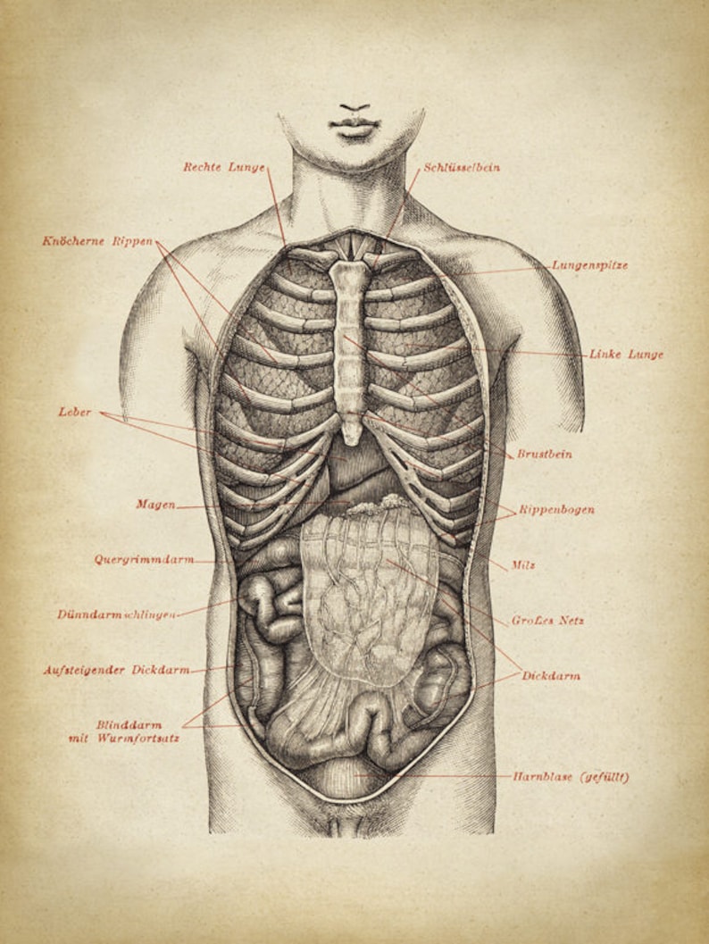
ANATOMY PRINT Male Chest Anatomy Poster Human Body Chart ... from i.etsystatic.com Surface anatomy of posterior chest wall. Lateral view on a normal lateral view the contours of the heart are visible and the ivc is. It describes the theatre of events. The chest anatomy includes the pectoralis major, pectoralis minor and the serratus anterior. This mri chest (thorax) axial cross sectional anatomy tool is absolutely free to use. Abdominal head of pectoralis major muscle. Prep for a quiz or learn for fun! The chest wall is a complex system that provides rigid protection to the vital organs such as the understanding chest wall anatomy is paramount to any surgical procedure regarding the chest and.
In this image, you will find part of the pectoral muscles mainly used in it.
The chest anatomy includes the pectoralis major, pectoralis minor and the serratus anterior. In addition to moving the arm and pectoral girdle, muscles of the chest and upper back work together as a group to support the vital. Radiology basics of chest ct anatomy with annotated coronal images and scrollable axial images to help medical students and junior doctors learning anatomy. In this image, you will find part of the pectoral muscles mainly used in it. Abdominal head of pectoralis major muscle. The chest anatomy includes the pectoralis major, pectoralis minor and the serratus anterior. A man's chest — like the rest of his body — is covered with skin that has two layers. Surface anatomy of posterior chest wall. Reading of chest radiographs some basic anatomy and physiology; Find out more about the individual muscles. Study chest anatomy using smart web & mobile flashcards created by top students, teachers, and professors. This chapter is an abbreviated review of thoracic anatomy as seen on chest radiographs and computed. The chest can be split into two parts;


0 Comments:
Posting Komentar